Difficulties in the diagnosis of neonatal hemangioma
Downloads
How to Cite
Bonifazi E. 2014. Difficulties in the diagnosis of neonatal hemangioma. Eur. J. Pediat. Dermatol. 24 (1): 49.
pp. 49
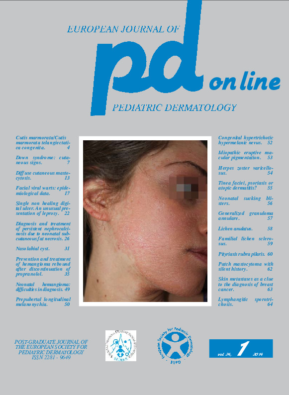
Abstract
A 20-day-old baby was first observed for the presence since birth of reddish lesions of the right hemiface. The physical examination showed flat red lesions on the right forehead-eyelid region and under the right ear (Fig. 1) leading to diagnose port-wine stain of the first and third trigeminal branch and to recommend 595 nm dye laser.When the child presented in the second month for the first session of dye laser, we noticed that the lesion of the first trigeminal branch had remained unchanged, while on the patch of the third trigeminal branch proliferative micropapules had appeared (Fig. 2); the dermoscopic observation of the red lesion under the ear showed irregularly dilated vessels with structures similar to microaneurysms, sometimes reminiscent of a rosary; a more careful observation in the crease behind the ear put in evidence an obvious hemangioma in plaque (Fig. 3). These clinical features led to the final diagnosis of port-wine stain of the first trigeminal branch associated with hemangioma under the right ear.
Keywords
Neonatal hemangiomas
