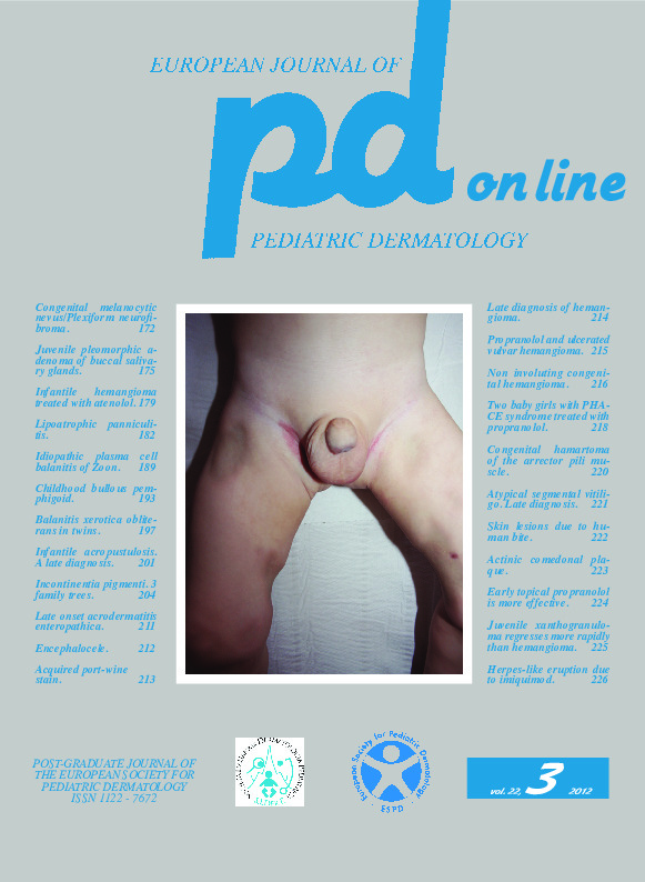
Encephalocele.
Downloads
How to Cite
Bufo R. 2012. Encephalocele. Eur. J. Pediat. Dermatol. 22 (3): 212.
pp. 212
Abstract
A 3-day-old infant was first observed due to the presence of a front swelling since birth. His family history was negative for similar lesions.Physical examination (Fig. 1, 2) showed on the frontal region in the mean level of the hairline a skin colored swelling of about 2 centimeters, with dilated follicular openings and hypertrichosis. Its consistency was soft elastic.
There was also on the nose paraphysiological sebaceous hyperplasia of the newborn.
An ultrasonography showed in the mass a tissue coming from the underlying suture. Cranial CT (Fig. 3) confirmed that the tissue inside the mass came from the brain structures and had the same density of the meninges. The clinical and imaging data led to the final diagnosis of cranial encephalocele so that the infant was addressed to neurosurgery.
Keywords
Encephalocele
