Late diagnosed nevus spilus.
Downloads
How to Cite
Garofalo L. 2011. Late diagnosed nevus spilus. Eur. J. Pediat. Dermatol. 21 (4): 245.
pp. 245
Abstract
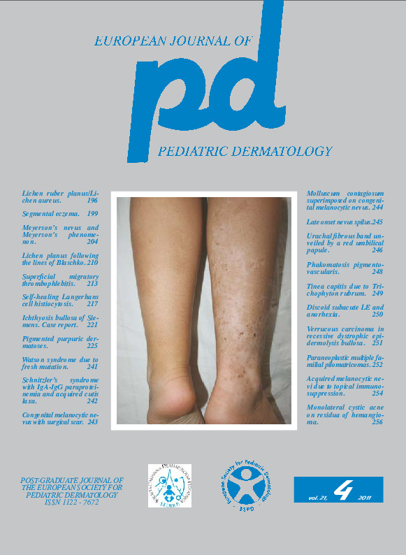
Case report. A 7-year-old boy showed hyperpigmented lesions of the back since birth. In the lumbar region there were 2 separate hyperpigmented patches: both had a uniform color, but the right patch was single with regular margins, while the left patch consisted of many small lesions close to each other (Fig. 1). On the back there were 3 acquired melanocytic nevi, two on left hemithorax and 1 on the hyperpigmented patch of the right lumbar region. These findings led to diagnose hypermelanic nevus on the right and dotted hypermelanic nevus on the left. After 4 years (Fig. 2) the two hyperpigmented lesions were unchanged, but about 15 acquired melanocytic nevi were visible, almost all on the right hyperpigmented patch, leading to the final diagnosis of nevus spilus. Dermoscopy of the right patch showed both at the age of 7 (Fig. 3) and 11 (Fig. 4) a reticular pattern with multifocal hypochromia.Keywords
Nevus spilus
