Lobulated pilomatricoma of the scalp.
Downloads
How to Cite
Garofalo L. 2011. Lobulated pilomatricoma of the scalp. Eur. J. Pediat. Dermatol. 21 (2): 125.
pp. 125
Abstract
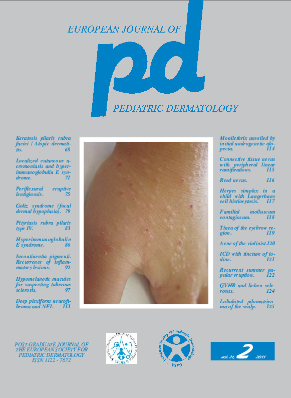
A 7-year-old girl known to us because in the past followed several times due to recurrent acute urticaria, came to our observation for the presence of a lesion of the scalp. Her mother reported that she had noticed the lesion from three months and during this time it had grown up to the current size. The lesion had never bled and was painless. Physical examination (Fig. 1) showed a roughly hemispherical neoformation of 5 mm, divided by septa into 6-7 lobules, of various color, bluish and in some places yellowish-pink. On palpation, the neoformation was hard, non tender, smooth-surfaced, movable on the deep layers. The lesion was excised under local anestesia and the histological examination showed a neoformation of basal cells and ghost cells with a giant-cell granulomatous infiltrate, leading to the final diagnosis of pilomatricoma.Keywords
Pilomatrixoma, Scalp
