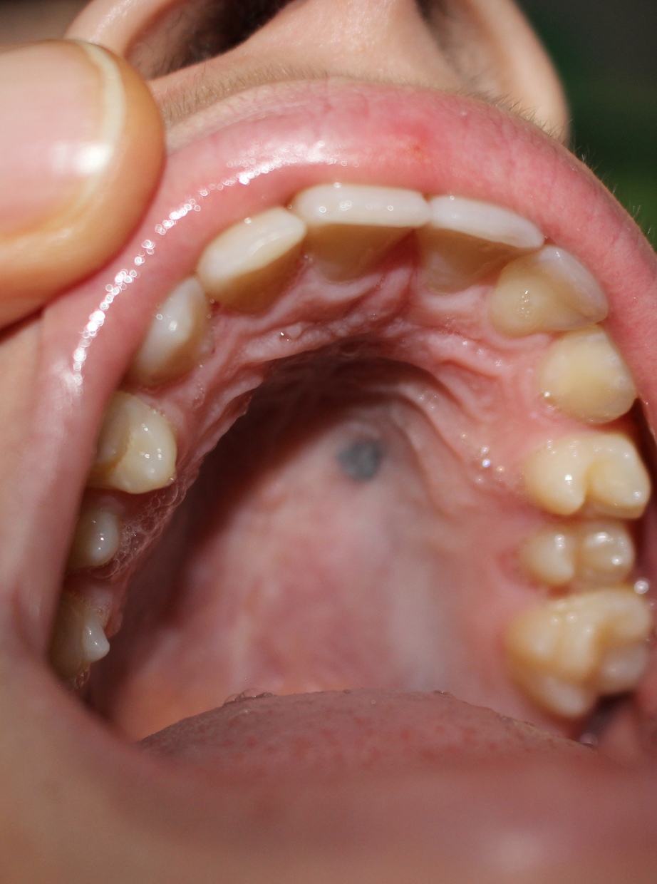
Blue nevus of the hard palate
Downloads
DOI:
https://doi.org/10.26326/2281-9649.31.2.2247How to Cite
Abstract
Blue nevus of the oral cavity is very rare. In particular, its frequency in our series of the last 50 years is 2 cases out of 100,000 pediatric skin disorders. Despite its rarity, it is the most frequent nevus after the intrachorionic nevus – equivalent to the intradermal nevus of the skin – among the melanocytic nevi of the oral cavity (3). Its diagnosis is based on the bluish color, which is due to the presence of melanin in the depth of the chorion thanks to Tyndall effect.
In children, a similar pigmentation can be due to amalgam and graphite tattoos (4), which can be ruled out quite easily based on history. For this reason and the difficulties of execution, it is not advisable to do a biopsy to ascertain the diagnosis of blue nevus, much less to exclude the hypothesis of a malignant blue nevus. In this regard, some Authors (5) argue that all melanocytic nevi of the oral cavity should be removed prophylactically due to the risk of malignant transformation linked to the constant chronic irritation of the oral mucosa during feeding, brushing of the teeth, etc. However, the larger case studies on melanocytic nevi of the oral cavity do not reach the same conclusions (3). The Authors of these studies reviewed 119 cases of melanocytic nevi of the oral cavity of the Dutch national registry collected in 25 years from 1980 to 2005; no patient had melanoma in an average observation period of 8.6 years.
These data, together with the rarity of malignant transformation in the blue nevus, the exceptionality of melanoma in pediatric age, and the non-existence in the literature of melanoma of the oral cavity under the age of 16 (6) suggest a conservative behavior in the case of blue nevus of the oral cavity in a child, unless the nevus continues to grow, becomes painful, bleeds, has irregular margins, or causes pigment dispersion into the surrounding mucosa (1). In other words, these conclusions are comparable to those adopted for longitudinal melanonychia of the child (2): the nevi of the oral cavity, of the nail matrix, or other particular sites must be biopsied following the same criteria for which a biopsy is performed in the common, acquired or congenital, melanocytic nevi of the skin in pediatric age.
