Hyperplasia of Tyson’s glands confirmed by polarized dermoscopy – The first report in a child.
Downloads
DOI:
https://doi.org/10.26326/2281-9649.30.3.2145How to Cite
Abstract
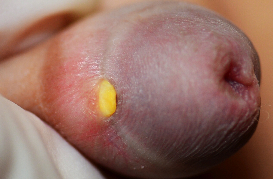
Tyson’s gland was first described by Edward Tyson (3). They are modified sebaceous glands on the coronal sulcus of the penis. They usually exist as a pair; multiple glands could be described as ectopic glands (5) or ectopic folliculosebaceous units (1). The importance of their role in secreting smegma is controversial.
They should be clearly differentiated from pearly penile papules, which do not secret smegma.
Normal Tyson’s glands might not be visible to naked eyes. When hyperplastic, they could be a cause of concern, and be brought to medical attention. In a study carried out in a clinic for sexually transmitted infections, hyperplasia of Tyson’s glands was the final diagnosis in 32/467 male patients attending the clinic (2). Hyperplasia is commonly reported in adults; however, it can occur in old age (4).
To our best knowledge, hyperplasia of the Tyson’s glands has never been reported in young children. Moreover, the polarized dermoscopic appearance of Tyson’s glands has not been demonstrated. We thus believe that it is worthwhile to report this child.
