Segmental elastic nevus with ipsilateral focal dermal hypoplasia.
Downloads
DOI:
https://doi.org/10.26326/2281-9649.29.4.2046How to Cite
Abstract
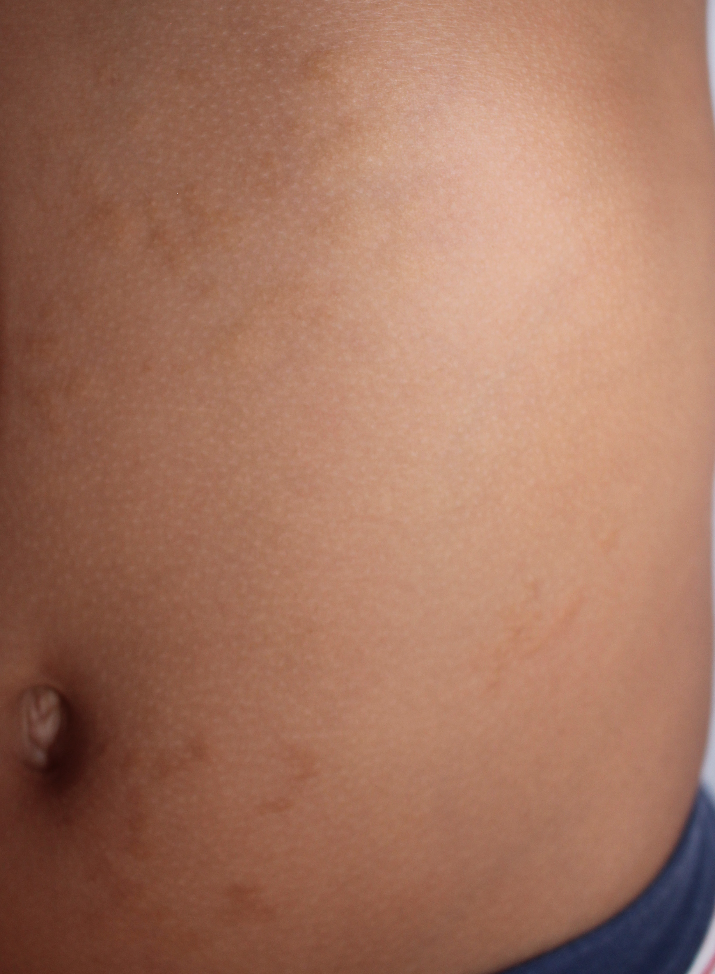
In the child you can rarely observe yellowish, scarcely visible but hard at palpation, infiltrative lesions, in the form of papules of a few mm or nodules and plaques. The lesions are histologically characterized by thickened and irregular elastic fibers. Their distribution can be asymmetric, segmental as in the current case (elastoma or elastic nevus) or extended and bilateral (dermatofibrosis lenticularis disseminata). These lesions can be associated with asymptomatic areas of increased opacity of long bones and pelvis that are important only because they can simulate bone metastases: this association is called Buschke-Ollendorf syndrome. The most extensive lesions can be linked to a mutation of the LEMD3 gene and transmitted as an autosomal dominant trait (1).
In the literature we have found only one case of segmental skin depressions associated with thick-ened elastic fibers (2).
