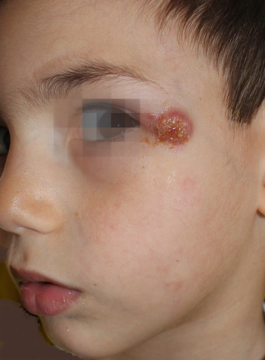
Lymphomatoid papulosis in a 5-year-old child.
Downloads
DOI:
https://doi.org/10.26326/2281-9649.29.3.2025How to Cite
Abstract
Lymphomatoid papulosis (PL) is a chronic-recurrent papulo-nodular skin disorder with histological appearance of malignant lymphoma, that behaves clinically as a benign self-healing disease, although the course can last many years. In the WHO-EORTC classification, PL is included together with large anaplastic cell lymphoma among CD30+ primary skin lymphoproliferative disorders. In 2016 Wieser et Al. (5) collected 251 cases in a population of children and adolescents with an average age of 9.3 years.
Clinically, PL is characterized by less than 2 cm in diameter, asymptomatic, more or less inflamed papulo-nodular lesions, that mainly affect the trunk, but also head and limbs. The lesions are usually few in number, counting and evolve characteristically in subintrant crops such as in pityriasis lichenoides. They start as papules, grow for about 2 weeks, can ulcerate and then regress spontaneously with transient pigmentary outcomes or scars in 1-2 months. The disease can evolve in months or more often in years, regressing spontaneously.
On the basis of the histological characteristics, immunohistochemistry and clonal rearrangement we distinguish different types, currently six. However, they do not differ substantially in clinical behavior. In type A, which is the most frequent - 80% of cases –, there are are large cells, sometimes similar to Sternberg cells, with lymphocytes, polymorphonuclear cells and eosinophils. Type B is characterized by epidermotropism resembling mycosis fungoides. In type C large CD30 + anaplastic cells prevail, making it similar to CD30+ large, anaplastic cell lymphoma. Type D is characterized by epidermotropism and variable amount of CD30 + cells, resembling primitive cutaneous CD8+ epidermotropic T-cell lymphoma (1). Type E is characterized by necrotic lesions and angioinvasive infiltrate (6). Type F is characterized by folliculotropism (2).
PL should be differentiated from pityriasis lichenoides that has smaller and more numerous, often purpuric lesions and is distributed in a centripetal or centrifugal manner; in doubtful cases histology shows the absence of CD30+ cells. More important is the differential diagnosis from CD30+ large anaplastic cell lymphoma that has a severe prognosis and needs specific therapy. Differentiation is easier clinically than histologically: clinically in lymphoma the nodules can be larger than 2 cm, continue to grow for more than 2-3 weeks and do not regress spontaneously; moreover, the number of lesions is usually lower in lymphoma.
Lymphomatoid papulosis may develop into lymphoma in 4% of cases but after 15 years (4). The clinical course in pediatric patients is chronic, sometimes with complete regression, but cases that have developed lymphoma in the adult, even extracutaneous after 40 years, are described though exceptionally (3).
