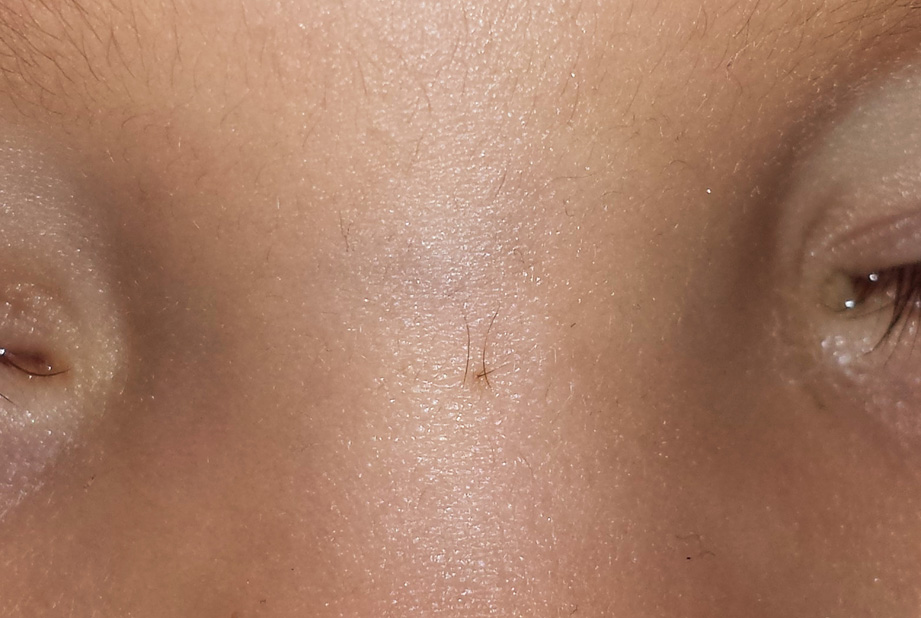
Closed nasal dermoid sinus.
Downloads
DOI:
https://doi.org/10.26326/2281-9649.29.3.2013How to Cite
Abstract
The nasal dermoid sinus is due to a closure defect of the neuroectodermal and mesodermal structures on the midline at the nasal level. During embryonic life the mesenchymal structures that will give rise to the frontal and nasal bone are formed by different centers which then merge and begin to ossify. Before these centers merge, between the glabellar region and the columella there are some spaces that play a significant role in the pathogenesis of the midline nasal defects: these spaces are the foramen caecum, which is located at the base of the frontal bone, the fronticulus frontalis between frontal and nasal bone and the prenasal space between nasal bone and cartilage.
During the fetal life an extension of the dura mater passes through the foramen caecum and comes into contact with the epidermis at the level of the fronticulus frontalis or of the prenasal space (Fig. 7, embryo).
When fusion and ossification occur the prolongation of the dura mater regresses, losing contact with the epidermis (Fig. 7, normal). When the physiological process of closure and ossification of these spaces is incomplete, it may happen that the epidermis penetrates deeply, moving towards the meningo-encephalic structures (Fig. 7, pathological) and that the latter remain in contact with the epidermis.
In milder cases, a fistulous path (dermoid sinus) is formed with an operculum at the epidermal level and a blind bottom at the level of the subcutaneous or osteocartilaginous structures; the dermoid sinus may end in a cystic dilation (dermoid cyst) or the cyst may not communicate with the epidermis (1, 2). The dermoid sinus and dermoid cysts in addition to the epidermis contain appendages (sebaceous glands, sweat glands and hairs). Clinically, you can see an operculum from which hairs, a serous liquid and sebum come out or a deep cyst. In more severe cases the fistulous path can reach the meningo-encephalic structures and these can be found in the skin, without communication with the cranial cavity (glioma) or with communication (meningo-encephalocele). In the case of cutaneous fistula infections can occur; in severe cases the latter may involve the bone and meningo-encephalic structures.
