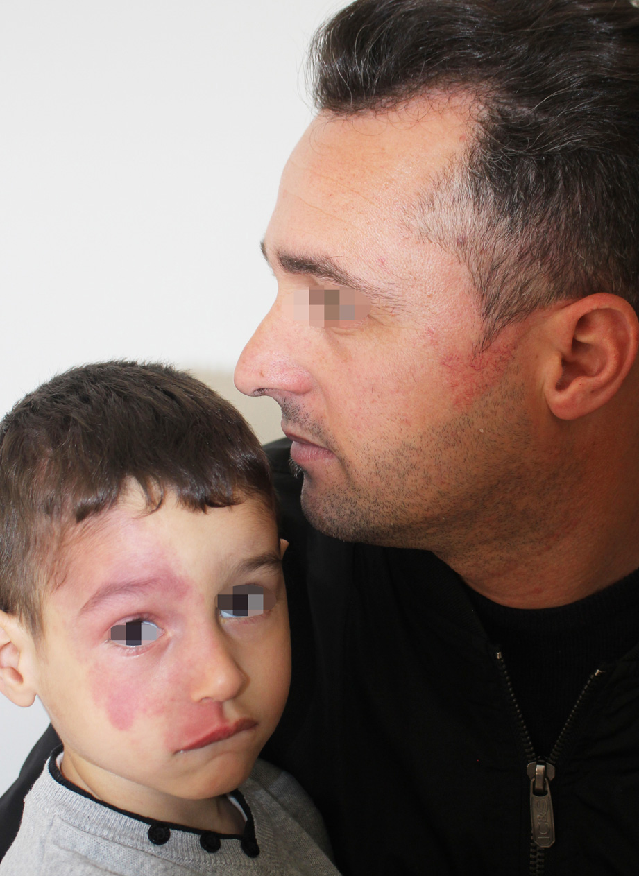
Port-wine stain present at birth in the child, acquired in the father.
Downloads
DOI:
https://doi.org/10.26326/2281-9649.29.2.1984How to Cite
Abstract
The current case lends itself well to the study of the relationship between port-wine stain, acquired port-wine stain and capillary malformation / arteriovenous malformation syndrome, both from an etiopathogenetic and differential diagnosis point of view.
It is known that in Sturge-Weber syndrome and congenital port wine stain somatic mutations of the GNAQ gene (7) can be found, but albeit rarely both in Parkes-Weber syndrome (4) and in Sturge-Weber syndrome (8) you can find RASA-1 mutations, which are detected much more frequently in the capillary malformation / arteriovenous malformation syndrome (3): the latter, presenting congenital and acquired red patches, may represent a trait d’union between congenital and acquired port-wine stain and must be differentiated from both. From a theoretical point of view it is however impossible to think that in our case the same mutation can be found in the father and the son; even indeed in the hypothesis of cutaneous and gonadal mosaicism in the father, the mutation in the child should be zygotic and therefore affect the whole organism.
As regards the differential diagnosis between these three vascular lesions, fundamental is the history that indicates the beginning at birth in the classic port-wine stain and the beginning after the birth in the acquired port-wine stain, while in the capillary malformation / arteriovenous malformation the lesions are both present at birth and acquired; the history also shows the familiarity in this last one unlike the other two.
The physical examination shows in the red patches of the capillary malformation / arteriovenous malformation a peripheral ischemic halo, a brownish nuance and sometimes hypertrichosis, in addition to possible signs of arteriovenous malformation; moreover the classic port-wine stain and the acquired one have a nevoid distribution, unlike the capillary malformation / arteriovenous malformation in which the distribution is random on the whole skin. Dermoscopy shows similar findingss in the classic port-wine stain and in the acquired one, ie dilated vessels regularly distributed (1); the presence of vessels of greater diameter, such as those present in the father of the current case, can be attributed to a greater depth of the lesion (5); in the red patches of the capillary malformation / arteriovenous malformation a brownish nuance and sometimes hypertrichosis can be seen (2).
