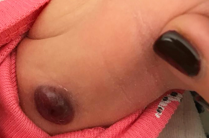
Congenital myofibroma in a newborn.
Downloads
DOI:
https://doi.org/10.26326/2281-9649.28.4.1934How to Cite
Abstract
Despite its rarity, myofibroma is the most frequent fibromatous neoformation of childhood. It can present in three clinical forms: isolated myofibroma, multiple-element myofibroma without visceral involvement and multiple-element myofibroma with visceral involvement. It is a neoformation typical of the child and when only the skin is affected in 50-60% of cases (4) is present at birth even in the premature (6). However, it can also be observed in adults, especially in the isolated form. It is a benign tumor and tends to regress spontaneously or does not relapses when it is removed provided that only the skin or bone is affected; when the internal organs, especially the lungs, are affected, myofibroma can behave destructively and lead to death; in fetal age also a large isolated cutaneous myofibroma can lead to death by anemization secondary to hemorrhage (1).
Histologically, myofibroma is characterized by a biphasic component with blunted nucleus cells reminiscent of myofibroblasts and roundish cells reminiscent of immature pericytes. According to Mentzel et Al. (8) myofibroma and infantile angiopericytoma belong to a broad spectrum of benign infantile myofibroblastic tumors and represent different stages of development of the same tumor; according to Zelger et Al. (9) this tumor after an initial monophasic stage acquires the classic biphasic appearance in which the lesion first becomes myoid, then fibrotic and finally sclerotic or hyalinized. (...).
