Characterization of melanocytes in the epidermis of human fetal scalp skin.
Downloads
DOI:
https://doi.org/10.26326/2281-9649.28.3.1870How to Cite
Abstract
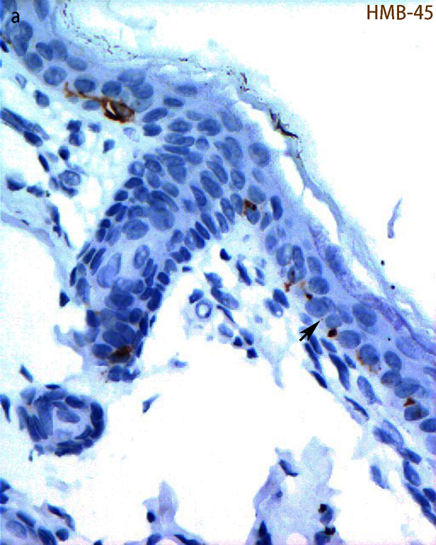
The study is aimed at observing melanocytes in fetal scalp epidermis, their dendrites and the ultrastructure of the melanin granules in those cells. To study the ultrastructure of melanocytes in human fetal interfollicular epidermis, we obtained a human fetal scalp specimen from an induced abortion of 140 to 150 days estimated gestational age. The scalp specimen was divided into two parts: one part was subjected to immunohistochemical staining with an anti-human HMB-45 antibody specific for melanosomes, while the other part underwent conventional preparation and staining for observation by transmission electron microscopy (TEM). Immunohistochemical analysis revealed that HMB-45 positive cells, namely melanocytes, were scattered at the base of the epidermal layer and in hair follicles especially within the outer root sheath. Analysis by TEM showed that melanocytes in the fetal scalp specimen had a clear cytoplasm and heterochromatic nuclei and contained various stages of melanosomes, most of them being immature (stages I-II). Furthermore, these cells contained abundant mitochondria, endoplasmic reticulum (ER), Golgi apparatus and ribosomes, as well as numerous cytoplasmic vacuoles. The dendrites from melanocytes that extended among keratinocytes contained many melanized melanosomes. However, the melanosomes were distributed in keratinocytes individually and sparsely. These observations revealed large numbers of immature melanosomes and a rich rough ER in epidermal melanocytes of the fetal scalp tissue compared to adult epidermis. Although the dendrites contained many mature melanosomes, few had been transferred to surrounding keratinocytes.
