Benign cephalic histiocytosis.
Downloads
How to Cite
Abstract
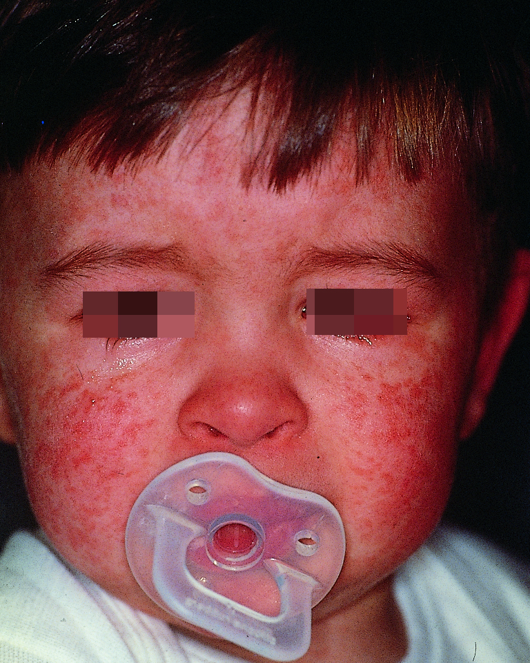
We report a 13-month-old boy with slightly elevated reddish-brown papules, measuring a few millimeters in diameter, scattered on the face, the neck and the shoulders. The eruption started at the age of 6 months on the cheeks and subsequently involved the forehead, the auricles and the scalp. Histological examination of a biopsy specimen from a papular lesion on the neck showed a well-circumscribed infiltrate in the superficial and mid-dermis consisting of large histiocytic cells with an irregular nucleus. Immunohistochemical studies carried out using an APAAP technique showed expression of CD11b and CD68 antigens on the histiocytic cells. The immunohistochemistry showed negative results for CD1a and S100 antigens. All laboratory investigations were within the range of normal values. Taken together, these findings suggested the final diagnosis of benign cephalic histiocytosis (BCH). This is a very rare non-Langethans cell proliferating histiocytosis, typically occurring in childhood.
