Congenital angiomatoid fibrous histiocytoma.
Downloads
DOI:
https://doi.org/10.26326/2281-9649.27.4.1514How to Cite
Abstract
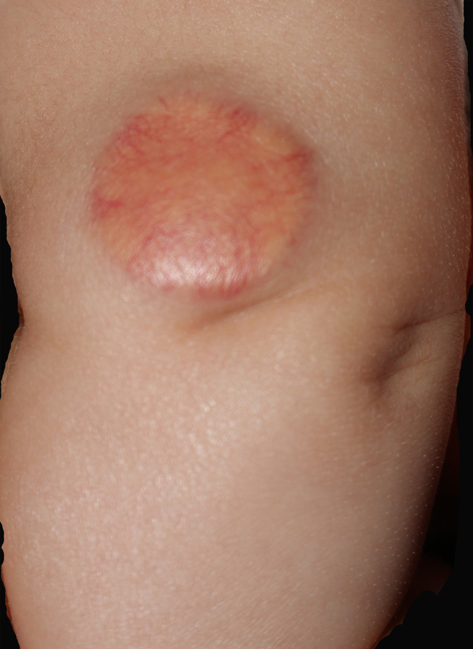
Angiomatoid fibrous histiocytoma is a rare low grade malignant tumor that affects the soft tissue mainly of the limbs in children and young adults.
Clinically, it is characterized by well defined nodules and plaques, with an average diameter of 2.5 cm (1) at the level of the limbs.
Histologically, it is characterized by nodules of spindle and oval histiocytoid cells, pseudo-vascular spaces filled with blood and surrounded by tumor cells, important lymphoplasmacytic cell infiltrate and a peripheral thick fibrous pseudocapsule.
In the initial case series of Enzinger (1), who first described the tumor as malignant angiomatoid fibrous histiocytoma, the incidence of mortality, metastasis and local recurrence was higher than it was later: at present (2) the incidence of local recurrences is 2 -11% and that of metastasis less than 1%.
